
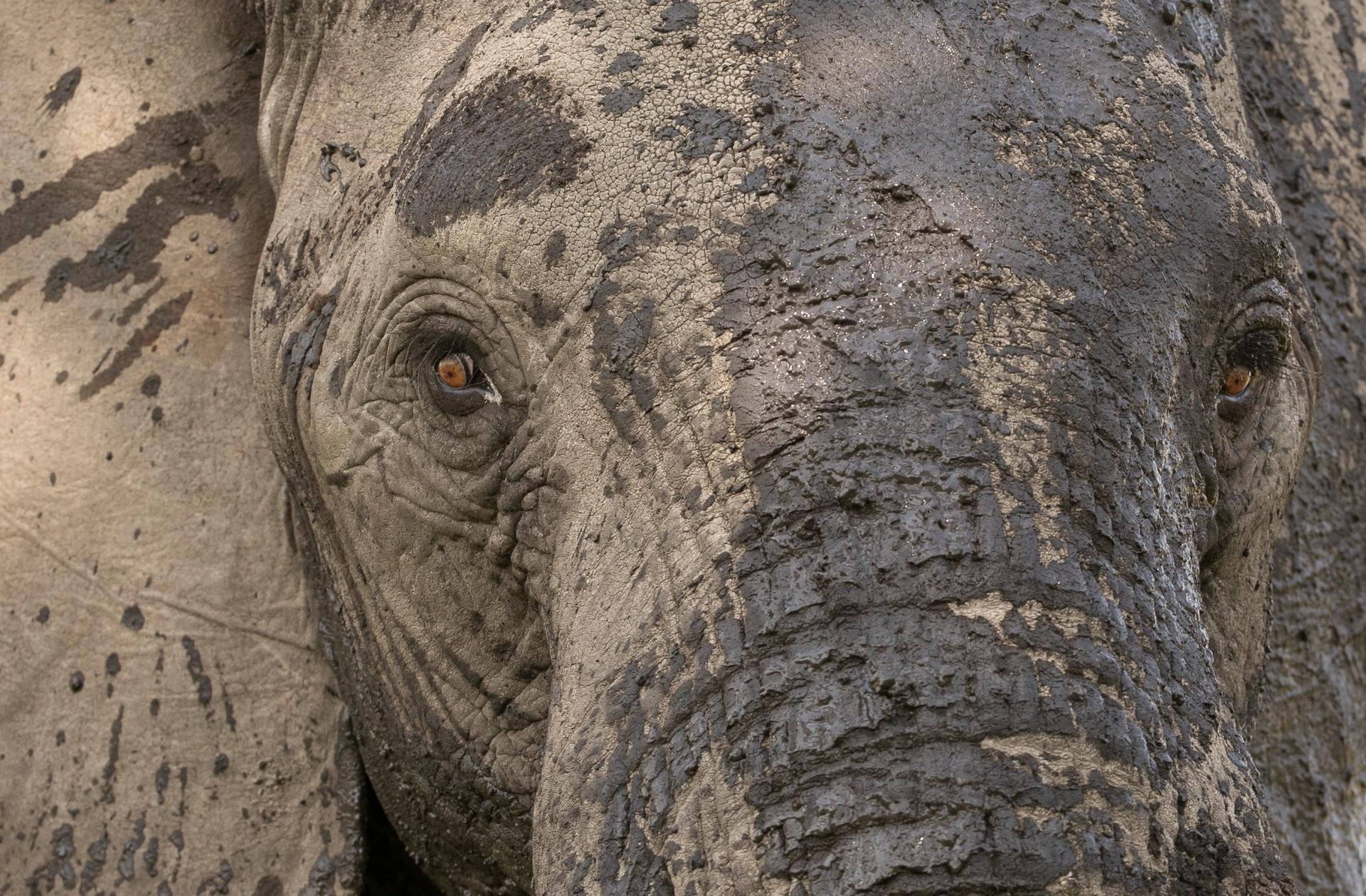
The elephant brain, at approximately 5,000 grams, is among the largest of any land mammal and exhibits striking structural complexity. Research demonstrates a highly expanded and intricately folded neocortex, an exceptionally large cerebellum, and well-developed sensory and emotional processing structures, including the amygdala and associated nuclei.
Encephalization quotient values place elephants within the range of great apes and some cetaceans, highlighting their substantial neural investment beyond basic bodily functions. Together these anatomical feature help explain elephants’ advanced cognitive and social abilities.

With a large body comes a large, complex brain. The elephant brain, at ~5,000 grams, is among the largest on the planet and is evolutionarily adapted to flourish in a complex, stimulating environment. Indeed, elephants and ~75% of cetacean species (i.e., whales and dolphins), along with humans and three pinniped species, belong to a small subset of species with brain masses >700 grams.
Until recently, very little was known about the elephant brain. There were only a few original articles specifically focused on the elephant central nervous system.
The brains of Asian (Elephas maximus) and African (Loxodonta africana) elephants rank among the highest for absolute and relative mass, and for cortical expansion. Their brains exhibit features comparable to those of some of the cetaceans and the great apes, including humans. The brain of a newly born elephant is approximately 50% of its adult weight, indicating a prolonged neurodevelopmental period wherein the environment will significantly shape the intricate neuronal microstructure.
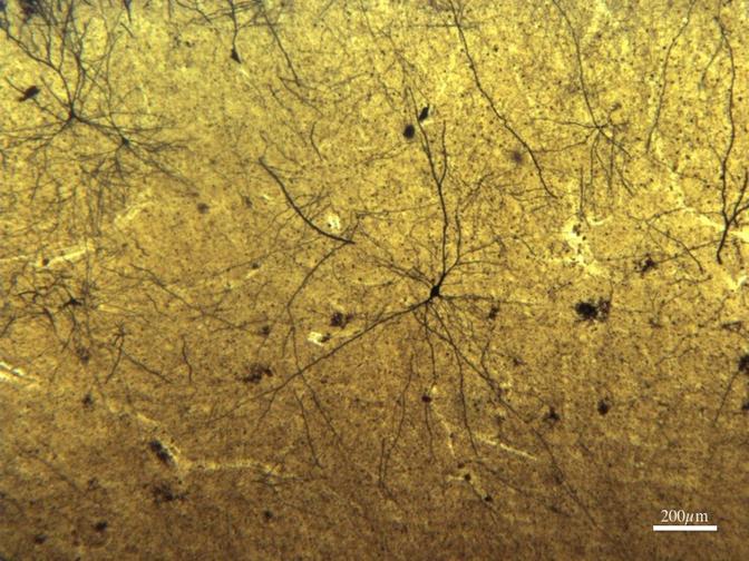
Golgi stain of elephant cortex under the microscope.
The cerebral cortex (or neocortex) is the outermost ribbon of nerve cells in the brain responsible for sensory elaboration and cognitive capacity (e.g., working memory, planning, decision making, spatial orientation, speech and language). Elephants have a large and highly convoluted neocortex, with the greatest volume available for cognitive processing of all land mammals. The volume of elephant neocortex relative to the rest of the brain ("neocortex ratio") has been shown to correlate closely with social group size, suggesting that it underwrites the cognitive skills needed for complex social living.
Moreover, the finding that neocortical ratio predicts the frequency with which primate species have been found to use tactical deception to solve social problems lends support to this argument. These cortical characteristics suggest a very elaborated sensory information/cognitive processing system.
The African elephant brain contains ~257 billion neurons, three times as many as the ~86 billion neurons in the adult human brain, with ~25 billion (or 97.5%) of these neurons in the cerebellum (compared to ~69 billion in the human) and only 5.6 billion in the neocortex (compared to 16.3 billion in the human). It should be noted that elephant neocortex appears to have a relatively low density of neurons, suggesting that the elephants' cognitive faculties may be concentrated in long-term processing and in synthesizing a great diversity of input over time.
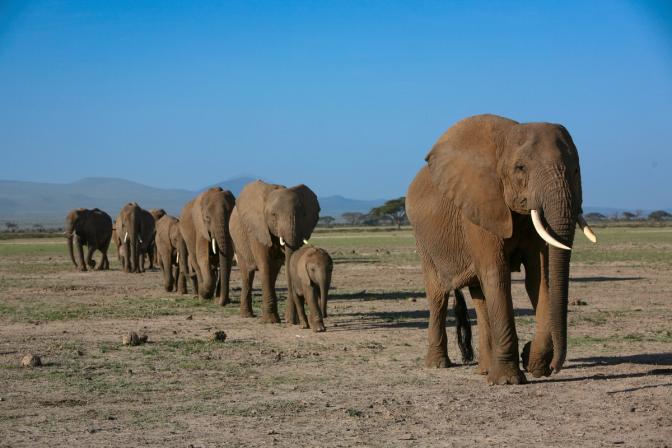
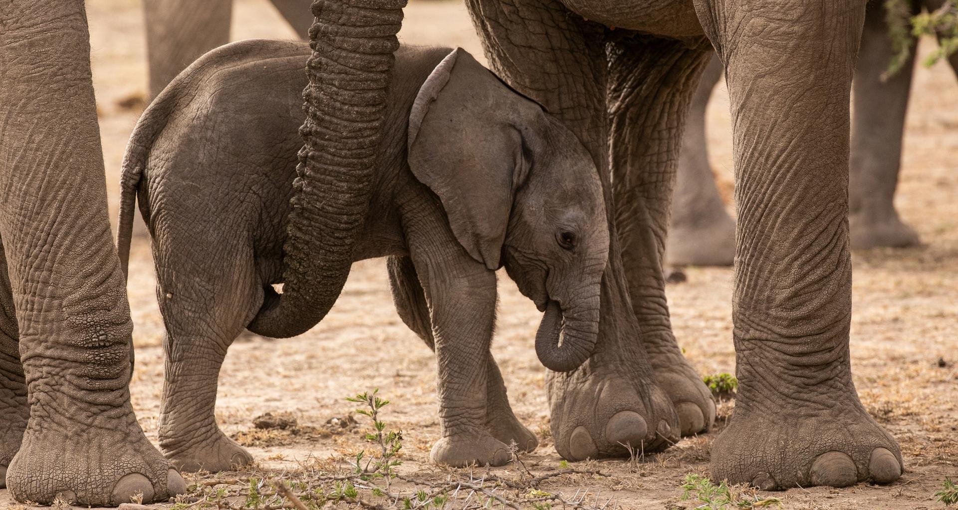
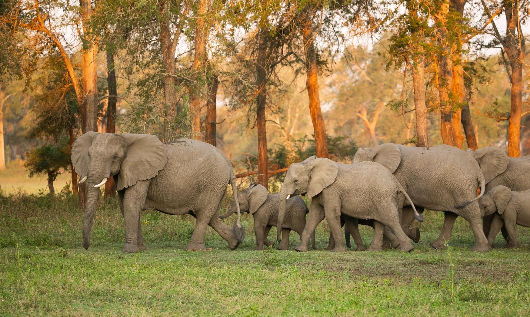
A procession of elephants in Amboseli - the large size of the temporal lobes and hippocampus may explain their prodigious memories and their ability to navigate over long distances.
One area of particular interest in the elephant is the temporal lobes and the underlying hippocampus, which are crucial to (declarative) memory functions, particularly of a spatial nature. An MRI study noted that elephants appear to have a somewhat large and highly convoluted hippocampus, although a more recent study by Patzke and coauthors suggests that the elephant hippocampus is about what one would expect in a brain of this size. Hakeem and coauthors estimated that the elephant hippocampus takes up 0.7% of the central structures of the brain in elephants, as compared to only 0.5% in humans.
Although the hippocampus is necessary for laying down new memories, the memories are not stored there. Instead, they are stored in the surrounding temporal lobes, which are particularly large and distinct in the elephant. The relative size of these two structures may explain their prodigious memories (for places, individuals, and events) and their ability to navigate over long distances.
An adult male and a young adult female illustrating sexual dimorphism in elephants
The corpus callosum is the main fiber tract that connects the two cerebral hemispheres. Although it has the largest absolute size recorded to date, the relative size compared to the rest of the brain is what one would expect in a brain of this size. The preliminary data also indicate a possible sexual dimorphism (females > males) in the size of the corpus callosum, which would suggest that elephants are the first non-primate to exhibit this sexual dimorphism.
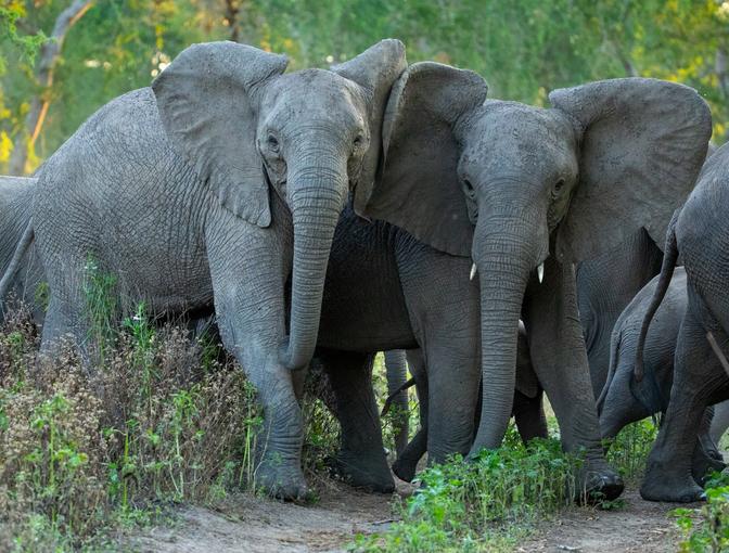
The amygdala is involved in evaluating incoming sensory information on an emotional level, especially with regard to the emotion of fear. Although the volume of the elephant amygdala is large, it is within the range of what one would expect in such a large brain. Nevertheless, the topological arrangement and relative size of some of the subnuclei (i.e., clustered groups of neurons that share the same function) in the elephant amygdala are unusual in comparison to other mammals. Subnuclei involved in the processing of emotional aspects of sensory information are particularly large; olfactory input appears to be very important in generating emotional states in the elephant.
The relatively large size of other amygdaloid subnuclei, in conjunction with the hippocampus, underscores the strong role that emotions play in memory formation. In an animal with such rich conspecific interactions, a relatively large amygdala and associated neural structures would be expected to play an essential for normal social functioning as well as for evaluating potential dangers in the environment.
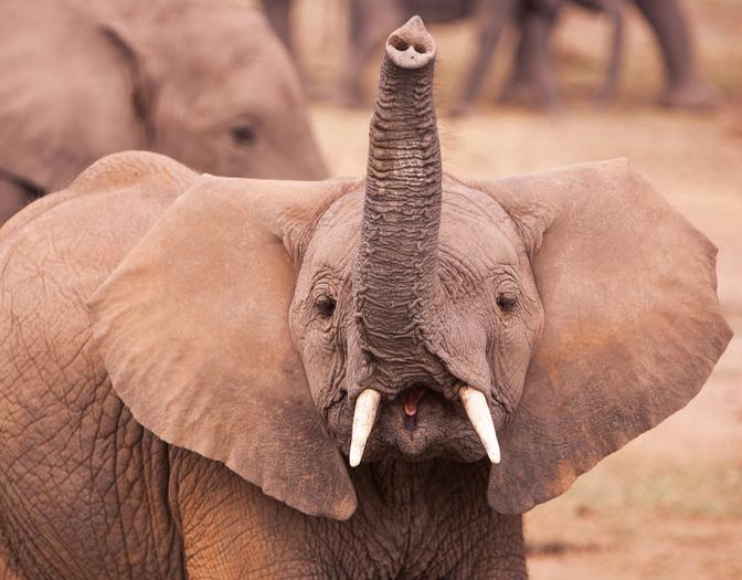
Although similar in many ways to what one observes in other mammals, there are many specializations (i.e., nuclei) in these subcortical structures. As one might expect, the nuclei involved in controlling movements of the trunk are quite elaborated. Brainstem regions involved in sound production and reception are also quite large, underscoring the fundamental importance of vocal communication in elephants.

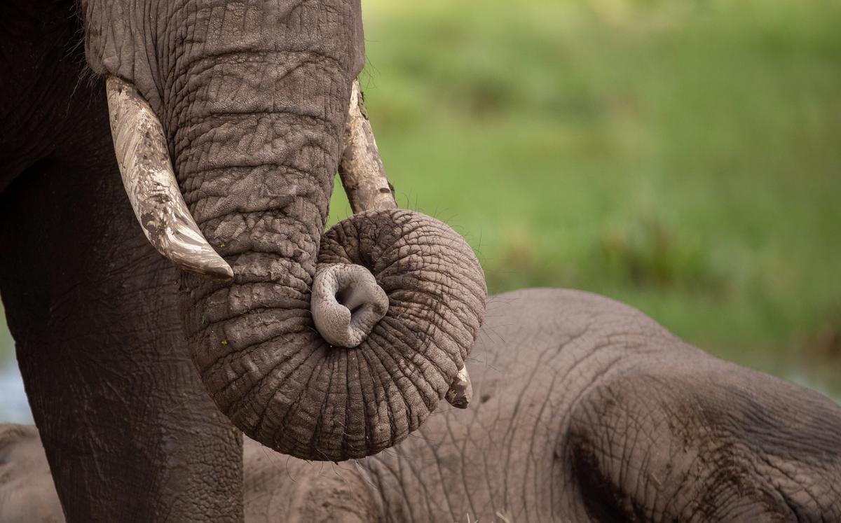
The trigeminal nerve is a cranial nerve that, among other functions, receives sensory information from the face and, in the elephant, crucially, from the trunk. In elephants, not surprisingly, this nerve and its associated ganglia, are extremely large. In fact, the nerve connections to the trunk are more substantial than the nerves to the rest of the elephant body. The sensory component of trunks is thus every bit as sensitive as the motor component is articulate.
Barasa A, Shochatovitz A. 1961. Grandezza e densità della cellule nervose della corteccia cerebrale de Elephas indicus. Rend Accad Naz Lincei. 30:246–249.
Bates LA, Poole JH, Byrne RW. 2007. Elephant cognition. Curr Biol. 17(16):R544–R546. https://doi.org/10.1016/j.cub.2008.04.019
Bower JM. 1997. Is the cerebellum sensory for motor’s sake, or motor for sensory’s sake? The view from the whiskers of a rat. Prog Brain Res. 114:463–497. https://doi.org/10.1016/S0079-6123(08)63381-6

The largest video and audio library of elephant behaviors.